

When dogs behave strangely, eat things they’re not supposed to (like Combat Roach Bait), or have visible irregularities, it can be worrying to us owners.
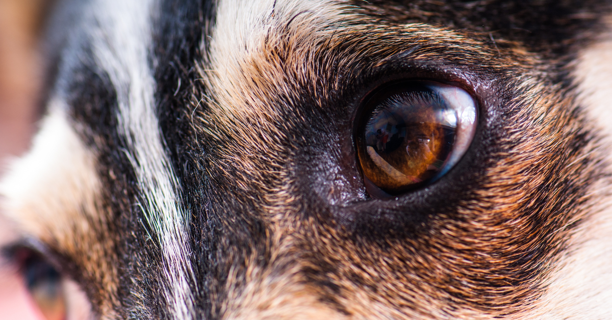
It can be especially concerning if there are abnormal structures in major parts of your dog’s body- like a brown spot on your dog’s eye.
If the spot does not change in size or color, and your dog is relatively young, this is likely to be natural pigmentation. Furthermore, older dogs can naturally get these pigmented spots due to a combination of genetics and environmental factors.
There are a few explanations for these brown spots, though typically the spot is caused by either pigmentary keratitis or ocular/eye melanoma.
Pigmentary keratitis involves the grouping of melanocytes, which are pigmented cells that give the spot its color in the eye.
They are harmless but the cause of pigmentary keratitis may need to be addressed. Further pigmentation can be stopped by administering the appropriate treatment relevant to the cause.
Eye melanoma involves a tumor in the eye which can be malignant (meaning it can spread and grow in size) or benign (meaning it only grows in size).
Malignant tumors are dangerous and urgent medical attention is required. Benign tumors are generally harmless, but they can grow to a size that puts pressure on local organs or structures. In this case, medical attention will also be required.
It is recommended to see the veterinarian or a veterinary ophthalmologist as there is a possibility that the brown spot could be a serious issue such as melanoma. The earlier your dog receives treatment, the easier it is to potentially cure the condition.
Brown/Black Spot On Dog’s Eye: Pigmentary Keratitis or Eye Melanoma?

To know whether the brown spot on your dog’s eye is caused by pigmentary keratitis or eye melanoma, you can check for general signs that are as follows:
Pigmentary Keratitis
- The Spot is Flat
- Located in the Sclera (White Portion of the Eye) or the Cornea (Transparent Window at the Front of the Eye)
- The Spot Roughly Stays the Same Size
- Your Dog is a Brachycephalic (Flat-faced) Breed e.g. Pug, Bulldog
Ocular/Eye Melanoma
- The Spot is Raised or Rounded
- The Spot has Relatively Distinct Borders
- Located in the Uvea (Iris, Choroid, Ciliary Body) or Limbus
- The Spot Increases in Size
- Eye Redness
- Dog Rubbing their Eyes
- Dog Breeds with Greater Skin Pigmentation e.g. Schnauzer, Golden Retriever
The list is not exhaustive, and your dog may only fulfil some, but not all conditions listed.
What is Pigmentary Keratitis on Dog Eyes?

Pigmentary Keratitis can appear as a dark brown blemish on the eye’s surface. The appearance is a result of a melanosome congregation. Melanosome contains melanin which produces the dark brown color that you see.
Pigmentary Keratitis is prevalent amongst brachycephalic dogs because of genetics and how their faces are structured. These include bulldogs, boxers, and pugs.
Causes of Pigmentary Keratitis
Pigmentary Keratitis can be caused by a combination of genetics and eye inflammation. Chronic eye inflammation is a major cause and leads to melanosome deposition in the cornea.
Eyelid diseases are a common cause of eye inflammation. Examples of eyelid diseases include eyelid tumors, irregular eyelashes, entropion (eyelid rolled inwards) and ectropion (eyelid rolled outwards).
Other causes of chronic eye inflammation include dry eyes, also known as Keratoconjunctivitis Sicca (associated with a lack of tear production) and irregular blink reflexes (leading to compromised ability to lubricate and protect the eye).
In some cases, eye inflammation is a result of corneal issues such as ulcers or caused by prior surgical procedures of the cornea.
Also Read: Vaseline For Dog Eye Boogers: Is it Safe to Use?
Clinical Signs of Pigmentary Keratitis
Pigmentation of the dog’s cornea is a clear sign of pigmentary keratitis. The appearance of the pigmentation ranges in a spectrum- from faint brown spots to darker blotches.
Owners will be able to see them relatively clearly under light although sometimes special equipment may be required to detect them.
You could use your hands to open their eyelids to detect pigmentation but you may accidentally poke them in the eye as dogs may be restless when doing so.
Diagnosis of Pigmentary Keratitis
A physical examination at your vet can determine if your dog has pigmentary keratitis. The vet will assess the condition of the cornea and look for other eye issues using tools such as an ophthalmoscope.
If your dog does have pigmentary keratitis, the vet will then try to determine the cause. The vet will examine for factors causing eye irritation such as the eyelids and the eye reflexes.
The vet may assess your dog’s tear production using the Schirmer Tear Test and dismiss the potential of corneal ulcers using a fluorescein eye stain. In more complex cases, your dog may be referred to an ophthalmologist.
Treatment for Pigmentary Keratitis
Treatment depends on the cause of your dog’s pigmentary keratitis. If the cause is identified and resolved, it could prevent further deposition of pigment in the cornea.
Surgery is often required to solve irregularities of the eyelid such as eyelid tumors, eyelash problems, entropion, and ectropion. Surgery helps fix the shape of the eyelid and prevents additional damage to the cornea.
If the cause is dry eye, medications that promote tear production will be administered to your dog. Artificial tears are also recommended to help lubricate your dog’s eye.
The pre-existing colored blotches are generally not removed via surgery as there are significant risks including blindness. Some medications may help reduce the visible spots although the effects are not the same for all dogs.
Currently, there are no treatments that can completely reverse pigmentary keratitis. The only exception would be dry eye whereby providing the appropriate tear film could fully remove the pigmentation.
Over time, the dark brown spots may become paler although they are unlikely to disappear.
What is Eye Melanoma in Dogs?
Eye melanoma, also known as ocular melanoma, is a type of cancer that develops from melanocytes proliferating uncontrollably in the eye.
Melanocytes are cells that produce melanin. Melanin is the pigment that makes hair, skin and eyes the color they are. Melanocytes are distributed throughout the body including the eyes.
Prominent areas of the eye where you can find melanocytes include the iris (the round structure surrounding the pupil and gives eyes its color) and behind the retina (the tissue layer at the back of the eye).
Eye melanoma typically is categorized as one of two types- uveal melanoma or limbal melanoma.
Uveal melanoma is the most prevalent type of tumor that occurs in the internal portion of the dog’s eye. Uveal tumors originate from the uvea which comprises the iris, choroid and ciliary body.
Around 80% of uveal tumors are benign and therefore will not spread to other organs.
Although 20% of uveal tumors are cancerous, the rate at which the cancer spreads is generally slower in dogs.
Limbal melanoma, also known as epibulbar melanoma, is benign (meaning it is generally harmless) and is rare.
Limbal melanomas occur at the limbus. The limbus is the barrier between the sclera (white portion of the eye) and the cornea (the outer transparent portion of the eye)
Cause of Eye Melanoma
The exact cause of eye melanoma is generally unknown in dogs. It could be a combination of environmental factors and genetics.
It is known, however, that eye melanoma is more likely to occur in breeds with more skin pigmentation. These include Schnauzers, Labrador Retrievers, Golden Retrievers and Cocker Spaniels.
Furthermore, eye melanoma is likely to develop as dogs grow into their middle to end stage in life. Although the typical age at which eye melanoma is diagnosed is at 9 years, dogs with a genetic predisposition for eye melanoma like some Labrador Retrievers may get it earlier at around the age of 1-2 years.
Signs of Tumor

Your dog’s eye will develop irregular structures and colors for both benign and malignant/cancerous tumors.
Uveal Melanoma
Uveal melanoma includes melanoma of the iris and melanoma of the ciliary body. As uveal melanoma develops, the pupil may become dilated and the pupil’s shape may change.
Other consequences of uveal melanoma include:
– Hyphemia (internal bleeding in the middle of the eye)
– Uveitis (inflammation in the middle of the eye)- leads to eyes becoming opaque
– Glaucoma (Elevated pressure in the eye)- the condition causes significant pain and leads to protruding eyes and potentially blindness. The pain from glaucoma can make your dog lethargic, squint, whine, and shake their heads.
Sign of Iris Melanoma – Single or multiple brownish-black spots on the iris which may be elevated or flat. Over time, the spots may develop and congregate, becoming an isolated and elevated mass.
Sign of Ciliary Body Melanoma – A dark mass in the eye that bulges out the pupil.
Limbal Melanoma
A primary sign of limbal melanoma is an elevated, brownish-black spot originating from the limbus’s boundary which spreads across the sclera (the white portion of the eye).
The melanoma typically also spreads to the cornea and causes corneal inflammation. This causes the cornea to become cloudy.
In cases where limbal melanoma spreads from the surface of the eye, conjunctivitis (where the eye’s surface becomes inflamed) and excess production of tears can occur.
Progression of Eye Melanoma
If your dog does not receive treatment, eye melanomas can lead to irregular eye structure, abnormal eye function and deterioration of eye health.
Uveal Melanomas
The rate of growth of uveal melanomas is slow.
Malignant uveal melanomas can spread to areas like the lungs, though this is rare.
Benign uveal melanomas will not spread to other regions but the tumor can expand and affect the structure and function of other eye structures as it grows. It can lead to eye rupturing and invasion of the eye socket.
If there is no treatment, long term consequences include glaucoma, uveitis, and impaired vision.
Limbal Melanomas
The rate of growth of most limbal melanomas is also slow. Although growth is slow, it can interfere with the function and shape of other eye structures including the cornea and causing corneal inflammation.
There have been no findings that suggest that limbal melanomas will spread to other regions.
Diagnosis of Eye Melanoma

Eye melanoma is diagnosed by assessing clinical signs and the tumor’s structure.
At the vet, your dog will undergo a series of examinations starting from a physical examination, then an eye assessment. Some of the tools used are as follows:
- Ophthalmoscope – examines the eye’s internal compartments
- Tonometer – estimates the internal pressure within the eye
- X-rays or Ultrasounds – to work out the tumor’s size, how far it may have spread and to rule out relatively harmless cysts.
Your dog may also undergo a process called staging. Staging is primarily used to assess the development of the cancer- notably uveal melanomas as 20% are malignant. Malignant means the tumor spreads outside of the eye.
The process involves various assessments including urinalysis (urine tests), bloodwork (blood tests) and sampling neighboring lymph nodes which helps determine how far the cancer has spread.
Treatment of Eye Melanoma
Uveal Melanoma
The method of dealing with uveal melanoma is dependent on factors including when it first appeared, how the uveal melanoma develops and the dog’s age and breed.
The mass is initially observed over a period of time to see how it develops. If it grows, the mass can be removed with no significant change in the eye structure and the dog’s vision.
Removal procedures include laser surgery or partial iridectomy (removing a portion of the iris via surgery).
In cases where the mass rapidly develops, the pupil’s shape is altered or there are signs of glaucoma, uveitis or hyphemia, the eye may need to be surgically removed.
The process of eye removal is known as enucleation and is recommended especially when uveal melanoma develops too fast or invades other local structures.
Prior to enucleation, the infected tissue is initially examined by a pathologist to see how active the tumor is. This will provide information on how far and how fast the tumor is likely to grow which will help the vet plan future actions where necessary.
If the tumor has spread or is beginning to, the vet will advise you to vaccinate your dog for melanoma and they will recommend using X-rays or ultrasound regularly to see if the tumor has spread further.
Currently, chemotherapy is not an option for treating eye melanoma as there is insufficient evidence that suggests it is effective.
Limbal Melanoma
Limbal Melanomas are initially monitored as they are benign and typically develop slowly.
Surgery will be required if the tumor invades other parts of the eye or develops too rapidly. Surgery involves removing the tumor and potentially parts of affected areas like the sclera or cornea.
Other treatments that may be used in conjunction with surgery includes radiation therapy, laser therapy or cryotherapy (where tissues are removed using extreme cold).
In severe cases where the tumor punctures the eye, enucleation is required. This process involves removal of the eye to get to the tumor to remove it.
Although enucleation can cure limbal melanoma, it does lead to blindness and is typically recommended only for severe and painful issues caused by the tumor.
In approximately 30% of dogs with limbal melanoma, the tumor will develop again, and further treatment will be needed.
Strange Behavior Linked to Eye Melanoma
If your dog does have eye melanoma, they will scratch or rub their eyes more often. Scratching or rubbing their eyes can cause eye infections and corneal ulcerations.
Infections and ulcerations can cause agonizing pain and lead to increased eye discharge, tearing, redness and abnormal eyelid movements such as squinting.
Enucleation (the removal of the eye) can seem like a poor choice, but it has the potential of stopping tumor-related pain and can save your dog’s life. In the long term, your dog should be able to smoothly adjust to the change in vision.
As tumors of eye melanoma are typically benign and are unlikely to spread, your dog is likely to recover well and live a healthy life after undergoing the appropriate treatment.
Conclusion
The brown spot in your dog’s eye could be a natural pigmentation that occurs with specific breeds or due to old age.
However, more often than not, the spot is typically explained by pigmentary keratitis or eye melanoma.
Pigmentary keratitis is generally caused by eye inflammation due to dry eyes, eyelid problems or abnormal eye reflexes. Treatment will depend on the cause and whilst treatment should stop further pigmentation, it will not reverse the brown spot in your dog’s eye.
Eye melanoma is generally caused by a combination of genetics and environmental factors. Treatment will focus on removing the tissue through surgery, laser therapy or other veterinarian-recommended procedures.
If you are ever uncertain, you should go to a veterinarian or a veterinary ophthalmologist to identify what the brown spot is. It can be relatively harmless or, it could be a severe issue like melanoma that can endanger a dog’s vision and life.

Heather Abraham is a professional blogger who owns two dogs, a cat, a parrot, and a leopard gecko. She has a connection with animals since she was a child. She shares her love for all pet breeds and provides information on pet food, toys, medications, beds, and everything else.
She is committed to learning about the internal workings of animals. Her work permits her to work closely with knowledgeable vets and obtain practical expertise in animal care. When she is not working, her love of animals continues in her writing. Her goal is to educate and uplift readers who also have a passion for animals through her writing.